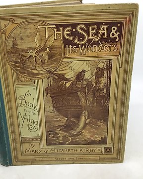
Mary and Elizabeth Kirby - The Sea and its wonders - 1890
Nº 84013345

Nº 84013345
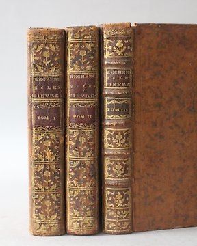
Samuel George Morton, M.D. - Illustrations of pulmonary consumption - Key & Biddle, 23 Minor Street, Philadelphia - Leather - 1834 - 183 pages (including index) - 24 cm x 15 cm.
This book is in good condition - Firm bindings - Small loss to spine - 19th century inscriptions previously marked over; otherwise all fine - Contains 12 full-page plates to the rear of the book, 11 of which are coloured (as called for), representing different diseases of the lungs - The plates are described as :
Plate 1.
Fig. 1 - A great number of tubercules in the apex of the hepatized lung, taken from a man aged twenty years, who died of pneumonia
Fig. 2 - Portion of the superior lobe of the right lung, converted into tuberculous matter, part of which is of a brownish red colour
Plate 2.
Fig. 1 - Part of the left lung of a man who died of consumption after five months illness
Fig. 2 - Angular crude tubercules disseminated in sound pulmonary tissue
Fig. 3 - Crude tubercules, some of which have suppurated at the margin
Plate 3.
Fig. 1 - Transverse section of an enormous encysted tubercle, (occupying nearly all of the superior lobe of the right lung), of a grayish yellow colour, and containing a few small abscesses
Fig. 2 - Portion of the left lung in a hepatized state, which, towards the right side of the figure, is passing into gray induration
Fig. 3 - Gelatinoid infiltration, of mixed olive and rose colour, with interspersed spots showing the transition of the infiltration into tubercular matter
Plate 4.
Fig. 1 - Angular crude tubercles and incipient abscesses, in a mass of engorged lung
Fig. 2- Funicular abscesses in the left lung; the bands of condensed, greenish tissue, are traversed by large ramifications of the pulmonary artery
Plate 5.
Fig. 1 - Portion of the right lung of a young man who died of phthisis (commencing with haemoptysis), after an illness of fourteen months
Fig. 2 - Section of the left lung, representing a large encysted abscess, the internal parietes of which are covered with red, extremely vascular granulations
Plate 6.
This plate represents a faithful transcript of the morbid appearances described in case 22.
Plate 7.
Fig. 1 - Superior lobe of the right lung, presenting several stages of gelatinoid infiltration, violet, gray and yellow, the latter being the crude state, or that which immediately precedes suppuration
Plate 8.
Fig. 1 - Profile view of John Little, drawn from life by my friend Dr. S. D. M'Neil, and showing deformity consequent to a dorsal tumour communicating with the lungs, and projecting over the interscapular region of the right side
Plate 9.
This plate represents a section of the superior lobe of the right lung, & c. of the above named John Little.
Plate 10.
Fig. 1 - The larynx, and part of the trachea, in a state if intense inflammation and ulceration, the cartilages being in many places ulcerated entirely through
Plate 11.
Fig. 1 - Portion of the pleura, partially covered with thick, and very firm adhesions, consequent to violent and long continued pleuritis
Fig. 2 - A portion of the tuberculosis lung, which, in consequence of partial pneumothorax, has receded from the ribs, but has been prevented from an entire collapse by two flattened cords, one passing from the upper, the other from the lower lobe, and uniting at the pleura costalis
Plate 12.
Fig. 1 - Portion of a tuberculosis lung, showing, immediately beneath the pleura, the remains of a small abscess which has become cicatrized and filled up with fibro-cartilaginous matter
Fig. 2- The bronchial mucous membrane in a state of intense inflammation (bronchitis), with small, whitish patches of ulceration
An exceptional book regarding 19th century medicine focussing on the diseases of the lungs.
Comment acheter sur Catawiki ?
1. Découvrez des objets d’exception
2. Faites la meilleure offre
3. Effectuez un paiement sécurisé