EBERT (Hermann). - [Super Sized Folio] Traité D’anatomie Pathologique Générale Et Spéciale - 1857-1861
![EBERT (Hermann). - [Super Sized Folio] Traité D’anatomie Pathologique Générale Et Spéciale - 1857-1861 #1.0](https://assets.catawiki.com/image/cw_ldp_l/plain/assets/catawiki/assets/2026/3/27/4/6/d/46dde198-eeec-4625-9ec8-7c30edd353ab.jpg)
![EBERT (Hermann). - [Super Sized Folio] Traité D’anatomie Pathologique Générale Et Spéciale - 1857-1861 #1.0](https://assets.catawiki.com/image/cw_ldp_l/plain/assets/catawiki/assets/2026/3/27/d/c/7/dc788a37-1fe8-4b6f-99ce-d6813293e022.jpg)
![EBERT (Hermann). - [Super Sized Folio] Traité D’anatomie Pathologique Générale Et Spéciale - 1857-1861 #2.1](https://assets.catawiki.com/image/cw_ldp_l/plain/assets/catawiki/assets/2026/3/27/f/3/3/f33716b4-8aea-41f4-9bdc-d097e4f85513.jpg)
![EBERT (Hermann). - [Super Sized Folio] Traité D’anatomie Pathologique Générale Et Spéciale - 1857-1861 #3.2](https://assets.catawiki.com/image/cw_ldp_l/plain/assets/catawiki/assets/2026/3/27/2/a/5/2a519550-48d9-4181-a0b3-5f75ad3739d6.jpg)
![EBERT (Hermann). - [Super Sized Folio] Traité D’anatomie Pathologique Générale Et Spéciale - 1857-1861 #4.3](https://assets.catawiki.com/image/cw_ldp_l/plain/assets/catawiki/assets/2026/3/27/a/1/7/a17d02ef-e341-47c7-a5c8-33810e98ab92.jpg)
Starting bid
€ 1
Add to your favourites to get an alert when the auction starts.

Catawiki Buyer Protection
Your payment’s safe with us until you receive your object.View details
Trustpilot 4.4 | 136750 reviews
Rated Excellent on Trustpilot.
Description from the seller
EBERT(ヘルマン)。 Traité d'anatomie pathologique generale et special ou description et iconographie pathologique des alterations morbides tant liquides que solides observees dans ls corps humain.
Text: [4], ii, 760; and [6], 733 [1] pp., including half titles, list of subscribers and index. Plates tissue guarded.
Plates: [2], 46; [2], 44 pp., with separate title-page to each part, 200 plates numbered I to CC, engraved after P. Lackerbauer, and engraved plates printed in colours and finished by hand.
Issued as a complete set of four large folio volumes, the publication consists of two volumes of text and two accompanying atlas volumes. The text volumes are extensive, totaling well over 1,400 pages, and provide systematic descriptions of pathological conditions affecting tissues, organs, and bodily fluids. The atlas volumes complement this scholarship with explanatory text and an extraordinary collection of engraved plates. Altogether, the work contains around 200 detailed illustrations, each designed to support and deepen the reader’s understanding of disease morphology.
What sets this treatise apart is the remarkable quality of its visual material. The plates, engraved on steel after the designs of the artist Pierre Lackerbauer, are meticulously hand-coloured, showcasing both macroscopic and microscopic pathological observations. These images were not merely decorative but served as essential scientific tools at a time when photography was not yet widely used in medicine. Renowned bibliographic references such as Garrison & Morton praises these engravings: “among the finest ever published, ” and we read: "The superb hand-coloured copperplate engravings of macro and micro-pathology in this work are among the finest ever published" -- Heirs of Hippocrates, 1803. See also A. Hisch & F. Hübotter. Biographisches Lexikon der hervorragenden Ärzte aller Zeiten und Völker (1962), Volume III: 706, and detailed study and praise in Edgar Goldschmid, Entwicklung und Bibliographie der Pathologisch-Anatomischen Abbildung (1925).
As a first edition, this work represents both a landmark in the history of pathological anatomy and a testament to the high standards of 19th-century medical publishing. It stands as a vivid reminder of a period when medicine, art, and craftsmanship intersected to produce works of lasting importance, bridging scientific inquiry with visual mastery.
HERMANN LEBERT (1813 – 1878) was a German physician and naturalist. After he received his medical doctorate (Zürich, 1834), he traveled throughout Switzerland, studying botany. For the next year and a half he studied in Paris, particularly under Baron Guillaume Dupuytren and Pierre Charles Alexandre Louis. In 1838 he settled in Bex, later changing between Bex and Paris. From 1842 to 1845 he worked mainly in comparative anatomy. In 1853 he accepted an invitation to become professor of clinical medicine in Zürich, and six years later he moved on to Breslau, where he held the same job. In 1862, he was elected as a member of the American Philosophical Society. In 1874 he returned to Bex, Switzerland, where he spent the rest of his life. Lebert was among the first to use the microscope in pathological anatomy, and thus contributed importantly to both pathology and clinical medicine. Traite d'Anatomie Pathologique (offered here) was Lebert's magnum opus. Combined with the superb detail of the illustrations by Peter Lackerbauer reproduced as copperplate engravings, it goes beyond Cruveilhier's famous Anatomie pathologique du corps humain (1829–1842) that includes only special pathology. The first volume contains a historical overview of pathological–anatomical iconography followed by a comprehensive essay on general pathology with relevant clinical case histories. The second volume is entirely dedicated to special pathology and also includes case histories and, among them, cases of tumor pathology. The scope of the two atlases is remarkable with the 200 large and mainly colored plates of the highest quality designed by outstanding artists such as the draftsman Lackerbauer and the engravers Oudet and Visto. The complete work was produced by the fine-art printers L. Martinet in Paris.
PETER LACKERBAUER (1823-1872). a prolific scientific illustrator of Swiss origin, who worked with Louis Pasteur, Claude Bernard and other scientists. More than twenty books have plates credited to Lackerbauer. For Pasteur he drew images of cells as seen in the field of a microscope. He also produced illustrations, including subtly varied tubes of wine under different conditions, for Pasteur's "Études sur le vin" (1866), and delicate colour images of the life cycle of silkworms for another major publication, "Études sur la maladie des vers à soie" (1870).
CONDITION REPORT
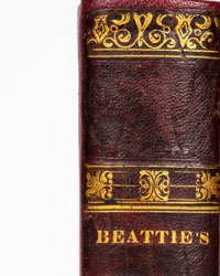
There is some scattered foxing (as seen in the last photos) and the binding are scuffed and lightly worn. But the bindings of these very large and heavy books are structurally sound. The scuffs are indicated in last photos.
EBERT(ヘルマン)。 Traité d'anatomie pathologique generale et special ou description et iconographie pathologique des alterations morbides tant liquides que solides observees dans ls corps humain.
Text: [4], ii, 760; and [6], 733 [1] pp., including half titles, list of subscribers and index. Plates tissue guarded.
Plates: [2], 46; [2], 44 pp., with separate title-page to each part, 200 plates numbered I to CC, engraved after P. Lackerbauer, and engraved plates printed in colours and finished by hand.
Issued as a complete set of four large folio volumes, the publication consists of two volumes of text and two accompanying atlas volumes. The text volumes are extensive, totaling well over 1,400 pages, and provide systematic descriptions of pathological conditions affecting tissues, organs, and bodily fluids. The atlas volumes complement this scholarship with explanatory text and an extraordinary collection of engraved plates. Altogether, the work contains around 200 detailed illustrations, each designed to support and deepen the reader’s understanding of disease morphology.
What sets this treatise apart is the remarkable quality of its visual material. The plates, engraved on steel after the designs of the artist Pierre Lackerbauer, are meticulously hand-coloured, showcasing both macroscopic and microscopic pathological observations. These images were not merely decorative but served as essential scientific tools at a time when photography was not yet widely used in medicine. Renowned bibliographic references such as Garrison & Morton praises these engravings: “among the finest ever published, ” and we read: "The superb hand-coloured copperplate engravings of macro and micro-pathology in this work are among the finest ever published" -- Heirs of Hippocrates, 1803. See also A. Hisch & F. Hübotter. Biographisches Lexikon der hervorragenden Ärzte aller Zeiten und Völker (1962), Volume III: 706, and detailed study and praise in Edgar Goldschmid, Entwicklung und Bibliographie der Pathologisch-Anatomischen Abbildung (1925).
As a first edition, this work represents both a landmark in the history of pathological anatomy and a testament to the high standards of 19th-century medical publishing. It stands as a vivid reminder of a period when medicine, art, and craftsmanship intersected to produce works of lasting importance, bridging scientific inquiry with visual mastery.
HERMANN LEBERT (1813 – 1878) was a German physician and naturalist. After he received his medical doctorate (Zürich, 1834), he traveled throughout Switzerland, studying botany. For the next year and a half he studied in Paris, particularly under Baron Guillaume Dupuytren and Pierre Charles Alexandre Louis. In 1838 he settled in Bex, later changing between Bex and Paris. From 1842 to 1845 he worked mainly in comparative anatomy. In 1853 he accepted an invitation to become professor of clinical medicine in Zürich, and six years later he moved on to Breslau, where he held the same job. In 1862, he was elected as a member of the American Philosophical Society. In 1874 he returned to Bex, Switzerland, where he spent the rest of his life. Lebert was among the first to use the microscope in pathological anatomy, and thus contributed importantly to both pathology and clinical medicine. Traite d'Anatomie Pathologique (offered here) was Lebert's magnum opus. Combined with the superb detail of the illustrations by Peter Lackerbauer reproduced as copperplate engravings, it goes beyond Cruveilhier's famous Anatomie pathologique du corps humain (1829–1842) that includes only special pathology. The first volume contains a historical overview of pathological–anatomical iconography followed by a comprehensive essay on general pathology with relevant clinical case histories. The second volume is entirely dedicated to special pathology and also includes case histories and, among them, cases of tumor pathology. The scope of the two atlases is remarkable with the 200 large and mainly colored plates of the highest quality designed by outstanding artists such as the draftsman Lackerbauer and the engravers Oudet and Visto. The complete work was produced by the fine-art printers L. Martinet in Paris.
PETER LACKERBAUER (1823-1872). a prolific scientific illustrator of Swiss origin, who worked with Louis Pasteur, Claude Bernard and other scientists. More than twenty books have plates credited to Lackerbauer. For Pasteur he drew images of cells as seen in the field of a microscope. He also produced illustrations, including subtly varied tubes of wine under different conditions, for Pasteur's "Études sur le vin" (1866), and delicate colour images of the life cycle of silkworms for another major publication, "Études sur la maladie des vers à soie" (1870).
CONDITION REPORT
There is some scattered foxing (as seen in the last photos) and the binding are scuffed and lightly worn. But the bindings of these very large and heavy books are structurally sound. The scuffs are indicated in last photos.
Details
書籍数
4
主題
Medicine
書籍タイトル
[Super Sized Folio] Traité D’anatomie Pathologique Générale Et Spéciale
著者/イラストレーター
EBERT (Hermann).
コンディション
Very good
最古の発行年
1857
最新の発行年
1861
高さ
46 cm
エディション
1st Edition
幅
30 cm
言語
French
原語
はい'
出版社
Paris: Baillière et Fils
装丁/素材
Half leather
付属品
Fold out maps or plates, Hand coloured illustrations, Tipped in plates
ページ数
1500
Sold by