Sir Charles Bell - The Anatomy of the Brain, Explained in a Series of Engravings. - 1802


Opens tomorrow
Starting bid
€ 1
Add to your favourites to get an alert when the auction starts.

Expert
Selected by Zena Chiara Masud
Holds a master’s degree in bibliography, with seven years of experience specialising in incunabula and Arabic manuscripts.
Estimate € 6,500 - € 7,200
Catawiki Buyer Protection
Your payment’s safe with us until you receive your object.View details
Trustpilot 4.4 | 123113 reviews
Rated Excellent on Trustpilot.
Description from the seller
Sir Charles Bell — The Anatomy of the Brain, Explained in a Series of Engravings. London, 1802. First Edition.
Bell, Sir Charles. The Anatomy of the Brain, Explained in a Series of Engravings.
London: C. Whittingham for T. N. Longman & O. Rees, and T. Cadell Jr. & W. Davies, 1802.
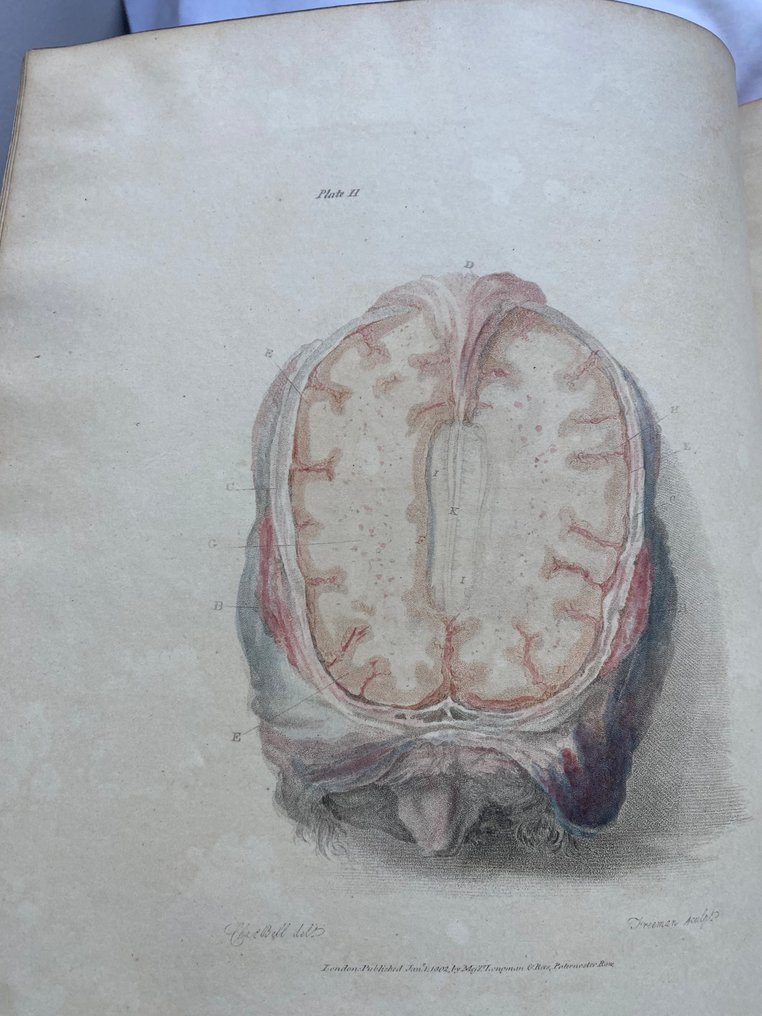
Small folio (29 × 24 cm). Title; pp. [iii]–vii; 87; 12 plates, 11 hand-coloured.
A LANDMARK IN NEUROANATOMY — EXCEPTIONALLY BRIGHT, WITH DISTINGUISHED PROVENANCE
The rare first edition of Sir Charles Bell’s most celebrated anatomical work, renowned for its union of scientific precision and artistic mastery. Bell—one of the most influential anatomists and surgeons of the early 19th century—personally drew the images from which Thomas Medland engraved the twelve aquatint plates. Eleven are beautifully hand-coloured, forming what historians have long regarded as the most exquisite visual record of early neuroanatomy.
Bell devoted his career to studying the nervous system, ultimately identifying the distinction between sensory and motor nerves, a discovery foundational to modern neuroscience. In this 1802 volume, his observational brilliance and draughtsmanship are on full display. Heirs of Hippocrates describes the plates as “probably Bell’s most beautiful work on neuroanatomy and one of the most beautifully illustrated in the entire literature,” while Garrison–Morton notes that Bell’s anatomical work “was the most important in the British Isles during the early part of the 19th century.” Plate I, in particular, is admired for its unprecedentedly accurate depiction of the cerebral gyri.
PROVENANCE
A period inscription on the flyleaf reads:
“Hune libruan yue in memorian servanduas Gulm. Mitchell M.D. L. Barlaw donavit Nov. 4th 1818.”
(“L. Barlaw donated this book to preserve the memory of William Mitchell, M.D., on November 4th, 1818.”)
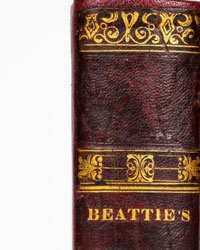
Later from the library of Kenneth Fitzpatrick Russell (1916–2006)—distinguished anatomist, medical historian, and author of British Anatomy 1525–1800. Russell regarded this copy as “a particularly fine copy of the rarest of all books by Bell.” It was rebound by Russell in handsome quarter red leather over red buckram.
Condition
A notably clean, crisp example.
Light damp-stain to upper right of title page; occasional spotting. Plates exceptionally vivid and fresh.
Binding tight and in very good condition.
Overall, an excellent, bright copy, exceedingly uncommon in such state.
RARITY AND MARKET NOTE
Copies of the 1802 first edition are scarce in commerce. The last notable listing, Bauman Rare Books (New York), offered a first edition in 2013 at $12,000; that copy has since been sold.
Sir Charles Bell — The Anatomy of the Brain, Explained in a Series of Engravings. London, 1802. First Edition.
Bell, Sir Charles. The Anatomy of the Brain, Explained in a Series of Engravings.
London: C. Whittingham for T. N. Longman & O. Rees, and T. Cadell Jr. & W. Davies, 1802.
Small folio (29 × 24 cm). Title; pp. [iii]–vii; 87; 12 plates, 11 hand-coloured.
A LANDMARK IN NEUROANATOMY — EXCEPTIONALLY BRIGHT, WITH DISTINGUISHED PROVENANCE
The rare first edition of Sir Charles Bell’s most celebrated anatomical work, renowned for its union of scientific precision and artistic mastery. Bell—one of the most influential anatomists and surgeons of the early 19th century—personally drew the images from which Thomas Medland engraved the twelve aquatint plates. Eleven are beautifully hand-coloured, forming what historians have long regarded as the most exquisite visual record of early neuroanatomy.
Bell devoted his career to studying the nervous system, ultimately identifying the distinction between sensory and motor nerves, a discovery foundational to modern neuroscience. In this 1802 volume, his observational brilliance and draughtsmanship are on full display. Heirs of Hippocrates describes the plates as “probably Bell’s most beautiful work on neuroanatomy and one of the most beautifully illustrated in the entire literature,” while Garrison–Morton notes that Bell’s anatomical work “was the most important in the British Isles during the early part of the 19th century.” Plate I, in particular, is admired for its unprecedentedly accurate depiction of the cerebral gyri.
PROVENANCE
A period inscription on the flyleaf reads:
“Hune libruan yue in memorian servanduas Gulm. Mitchell M.D. L. Barlaw donavit Nov. 4th 1818.”
(“L. Barlaw donated this book to preserve the memory of William Mitchell, M.D., on November 4th, 1818.”)
Later from the library of Kenneth Fitzpatrick Russell (1916–2006)—distinguished anatomist, medical historian, and author of British Anatomy 1525–1800. Russell regarded this copy as “a particularly fine copy of the rarest of all books by Bell.” It was rebound by Russell in handsome quarter red leather over red buckram.
Condition
A notably clean, crisp example.
Light damp-stain to upper right of title page; occasional spotting. Plates exceptionally vivid and fresh.
Binding tight and in very good condition.
Overall, an excellent, bright copy, exceedingly uncommon in such state.
RARITY AND MARKET NOTE
Copies of the 1802 first edition are scarce in commerce. The last notable listing, Bauman Rare Books (New York), offered a first edition in 2013 at $12,000; that copy has since been sold.
Details
Number of Books
1
Subject
Medicine
Book Title
The Anatomy of the Brain, Explained in a Series of Engravings.
Author/ Illustrator
Sir Charles Bell
Condition
Very good
Publication year oldest item
1802
Height
29 cm
Edition
1st Edition
Width
24 cm
Language
English
Original language
Yes
Publisher
London: C Whittingham, Dean-Street Fetter-Lane, for T. N. Longman and O. Rees, Paternoster-Row, and
Binding/ Material
Half leather
Extras
Hand coloured illustrations, Tipped in plates
Number of pages
100
Sold by